Read at The Van Gogh House inside the museum · the same label the museum panel shows, from content/inception/museum-labels/van-gogh-color-science.html

Art, chemistry, and time

The Violet That Time Hid

How science helps us imagine Van Gogh's original colours

The blue you see today may not be the blue Van Gogh painted.

In May 1889, soon after entering the hospital at Saint-Rémy, Vincent van Gogh painted the irises growing in its garden. Most of the flowers now appear blue. Research by the Getty Museum and Getty Conservation Institute found evidence that many of them were originally a deeper violet.
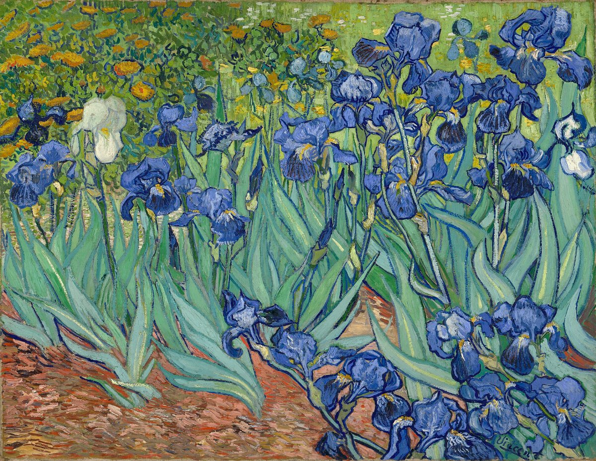
Vincent van Gogh's Irises: a dense bed of blue iris flowers with long blue-green leaves, an orange earth path, and a band of yellow-orange marigolds along the top.
Vincent van Gogh, Irises, 1889 · J. Paul Getty Museum, Los Angeles. The painting as it appears today. This is the present state of the work, not a reconstruction.

Where did the violet go?

To mix violet, Van Gogh added a red made from dye (called a “red lake”) to blue paint. The blue was tough. That red was not — light slowly bleached it. After more than a hundred years, much of the red is gone, so today our eyes mostly see the blue that was left behind.

The painting was not replaced. Its materials changed with time.
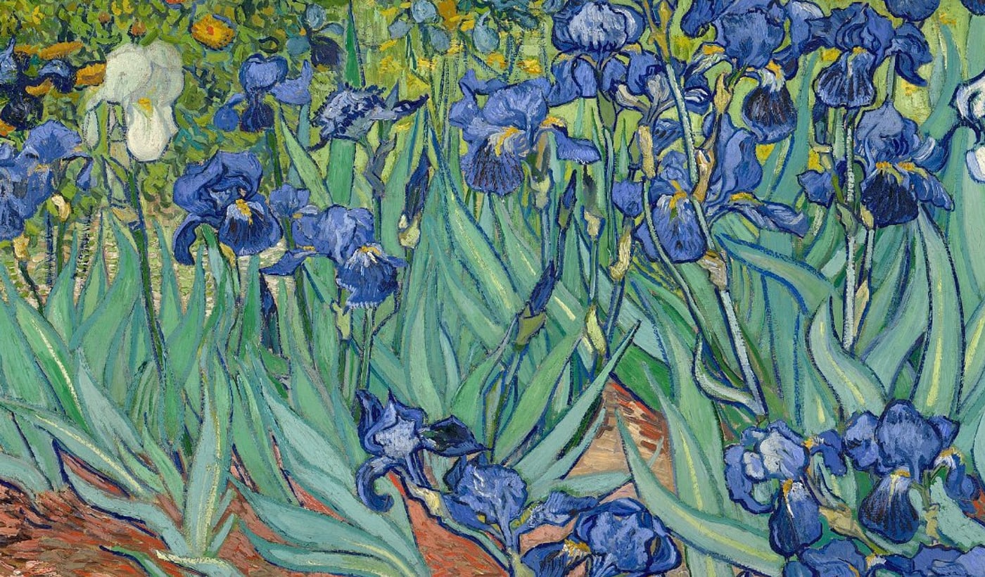
A close view of several iris flowers in the painting, showing blue petals built from short, separate brushstrokes against green leaves.
A direct crop of the public-domain painting, enlarged. These are the flowers that read as blue today.
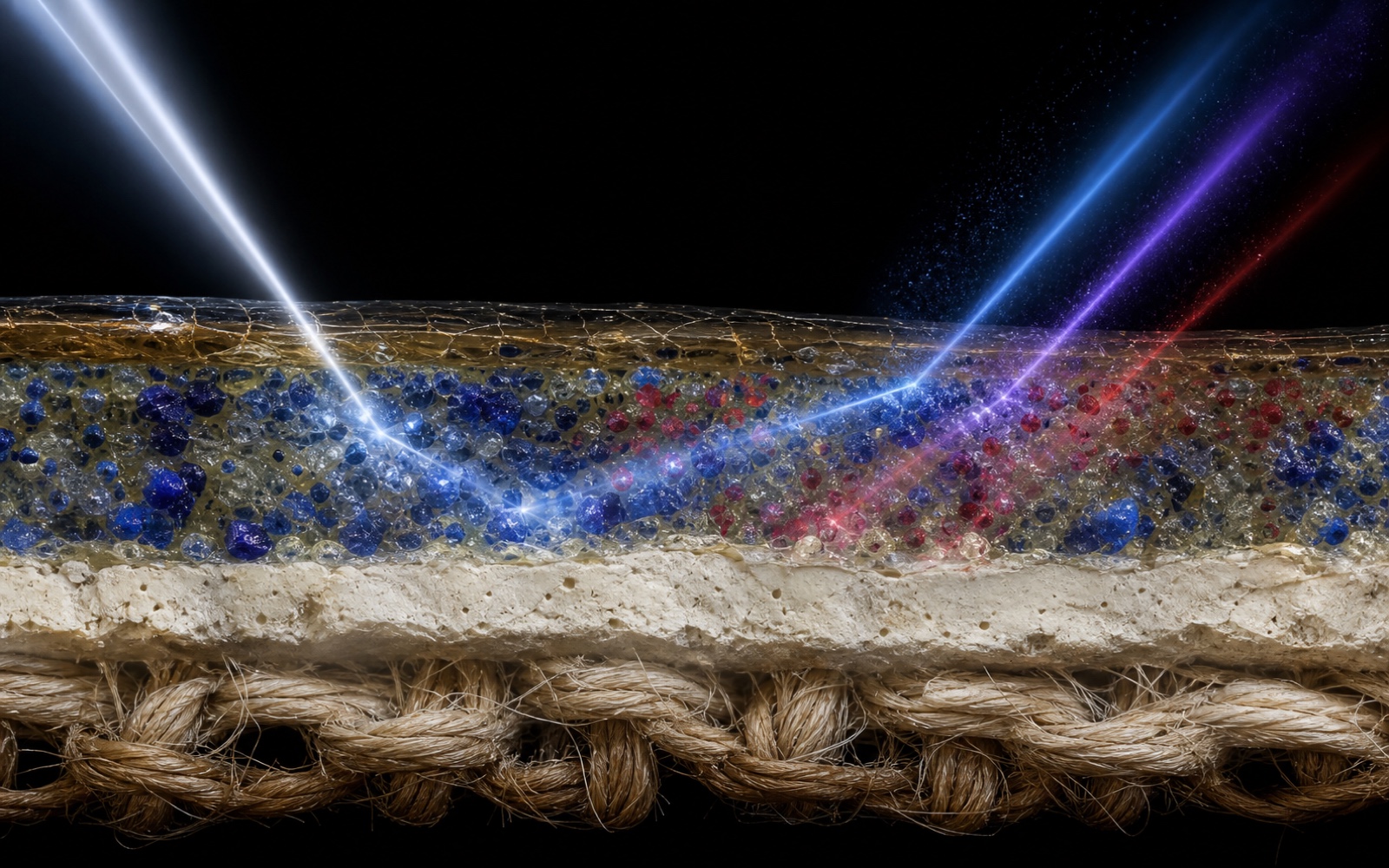
A cinematic microscopic cutaway through an intact painting, showing woven canvas, a pale ground, blue and red pigment particles in binder, aged varnish, and light scattering through the layers.
Concept visualization: an intact paint-layer model showing blue and red components, the pale ground, canvas, and the way light may scatter through them. It is an explanatory rendering, not a real paint sample and not a micrograph.

How can scientists find a colour that has faded?

No single instrument supplies the answer. Researchers compare several kinds of evidence.

  1. Looking through a magnifier. One blue iris has a tiny chipped spot — and inside it, protected under the surface, the paint is still purple.
  2. An element-mapping scanner, called XRF. Without touching the painting, it maps which chemical elements sit where. The faded red paint contained an element called bromine — so the bromine map shows where that red used to be. The scanner cannot photograph the lost colour itself; it finds chemical fingerprints, and scientists work out the pigments from them.
  3. Light tests and chemistry tests. Several instruments — reflectance spectroscopy, Raman spectroscopy, gas chromatography–mass spectrometry — shine light on the paint or study tiny samples of it, each reading a different kind of fingerprint, to work out which pigments and which glues the paint was made from.
  4. Microfade testing — the tiny-fade test. Scientists shine a pinpoint of strong light on a spot almost too small to see, to check which colours would still fade — so the museum knows how gentle its lights must be.
  5. Photogrammetry — 3D photography. Very sharp photos, taken from many angles, record not just the colours but the little ridges each brushstroke left in the paint.
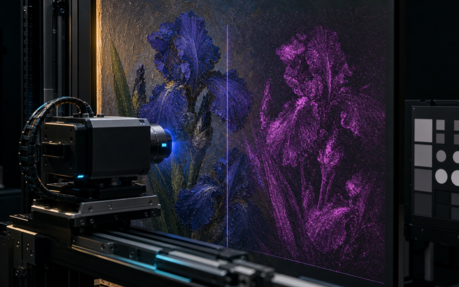
A non-contact XRF scanner moves on precision rails in front of blue-violet painted irises while a registered magenta density field visualizes where a selected element might be distributed.
Schematic element map — not Getty measurement data. This concept visualization shows how a non-contact XRF scan can map an element's distribution across the same flower shapes.
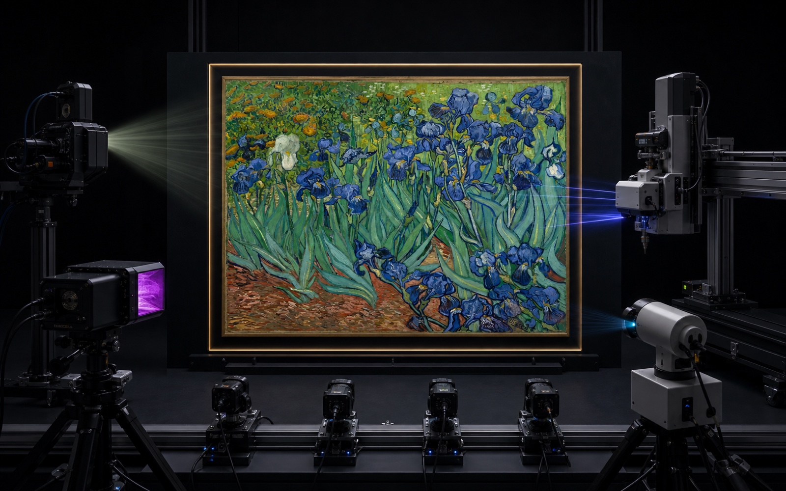
Van Gogh's Irises stands in a dark conservation laboratory, surrounded at a safe distance by cameras, optical sensors, a scanning gantry, and raking-light equipment.
Concept visualization: several independent, non-contact methods converging on the same painting. Agreement between them is what makes a conclusion strong.

From evidence to a digital reconstruction

First the researchers find every area where the paint changed, and sort them by how much each one changed. Then they adjust the colour bit by bit, following each brushstroke and each paint mixture — instead of dropping one purple filter over the whole picture.
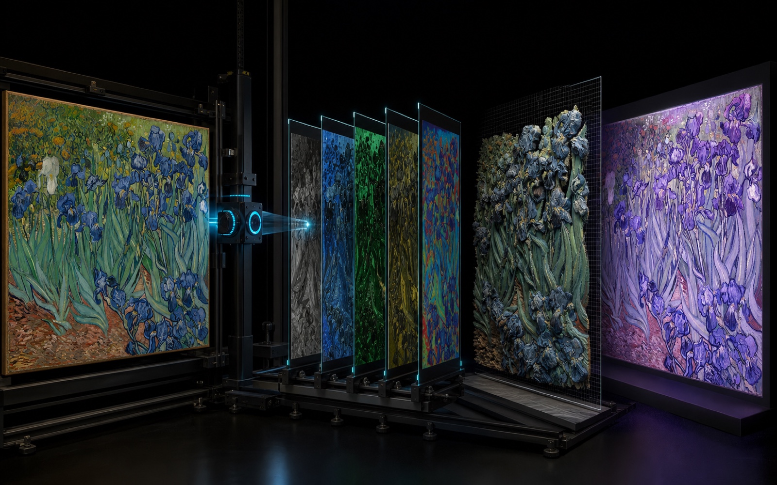
Van Gogh's Irises appears unchanged at the left of a dark imaging system; registered spectral planes lead to a three-dimensional brushstroke model and a separate violet digital hypothesis at the right.
Concept visualization of the workflow: present appearance, several registered evidence layers, a 3D digital twin, and a colour-adjusted hypothesis kept visibly separate from the original.

Getty even put the corrected colours onto a 3D model of the painting's bumpy surface, and 3D-printed it — because colour is not the only thing your eyes read. Light sliding sideways across a raised brushstroke changes how that colour looks.
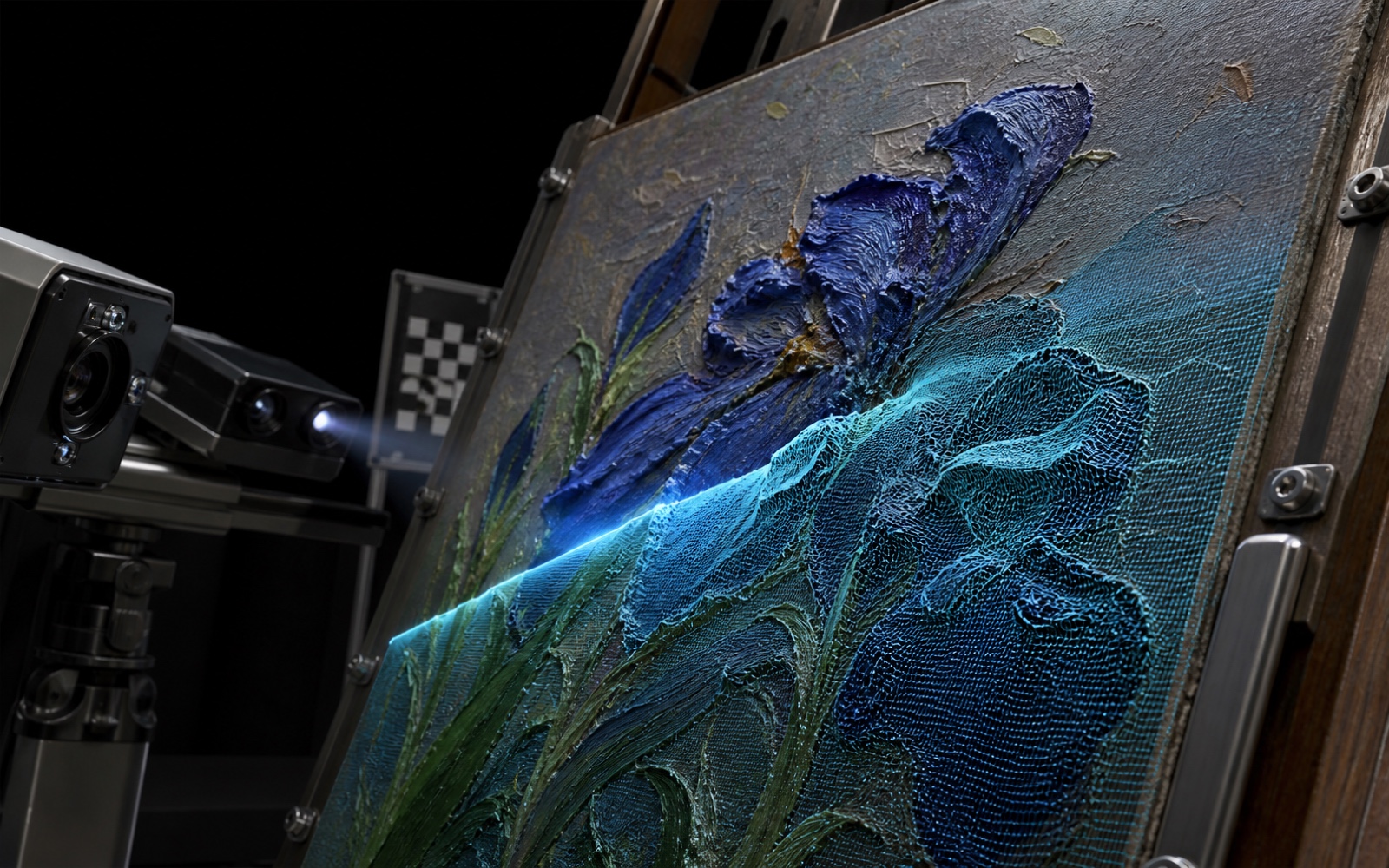
A structured-light scanner records thick blue iris brushstrokes while a precisely registered cyan mesh takes shape over the lower half of the painted surface.
Concept visualization of non-contact 3D capture: a registered mesh follows the relief of raised brushstrokes. It is an explanatory rendering, not a scan of Getty's 3D model.
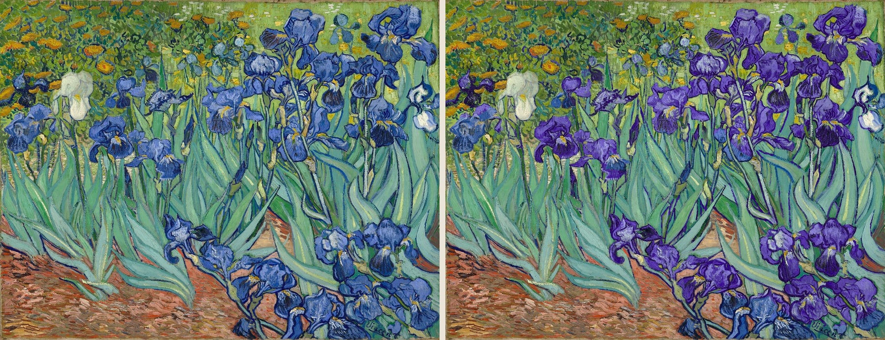
The Irises painting shown twice side by side. In the left copy the flowers are blue; in the right copy the same flowers are violet, while leaves, path, and marigolds are unchanged.
Left: the public-domain painting as it appears today. Right: an evidence-constrained colour-direction hypothesis that moves selected blue iris paint toward violet. This is a scientific approximation, not Getty's calibrated reconstruction and not a change to the original painting.

This side-by-side is not time travel, and it is not the only possible answer. It is a visual hypothesis constrained by evidence. The original painting remains untouched.

A second clue: colours protected by a frame

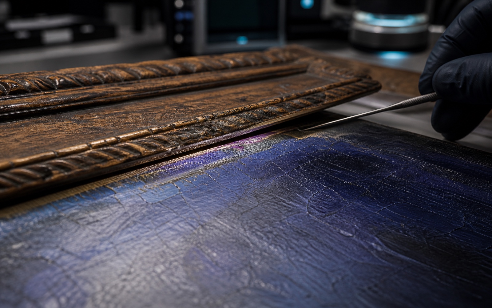
Colour evidence can also survive at the edge of a painting. Areas covered by a frame receive less light and collect less surface dirt. In studies of Van Gogh's The Bedroom, faded red lake pigments helped turn originally purple walls toward blue and a pinker floor toward brown.

Today, researchers can compare the protected strips with the faded parts, then teach a computer program to shift colours using clues from the painting itself. Helpful — but the program needs an expert watching it. A program that only learns from what it can see might “fix” places where the paint never actually changed.
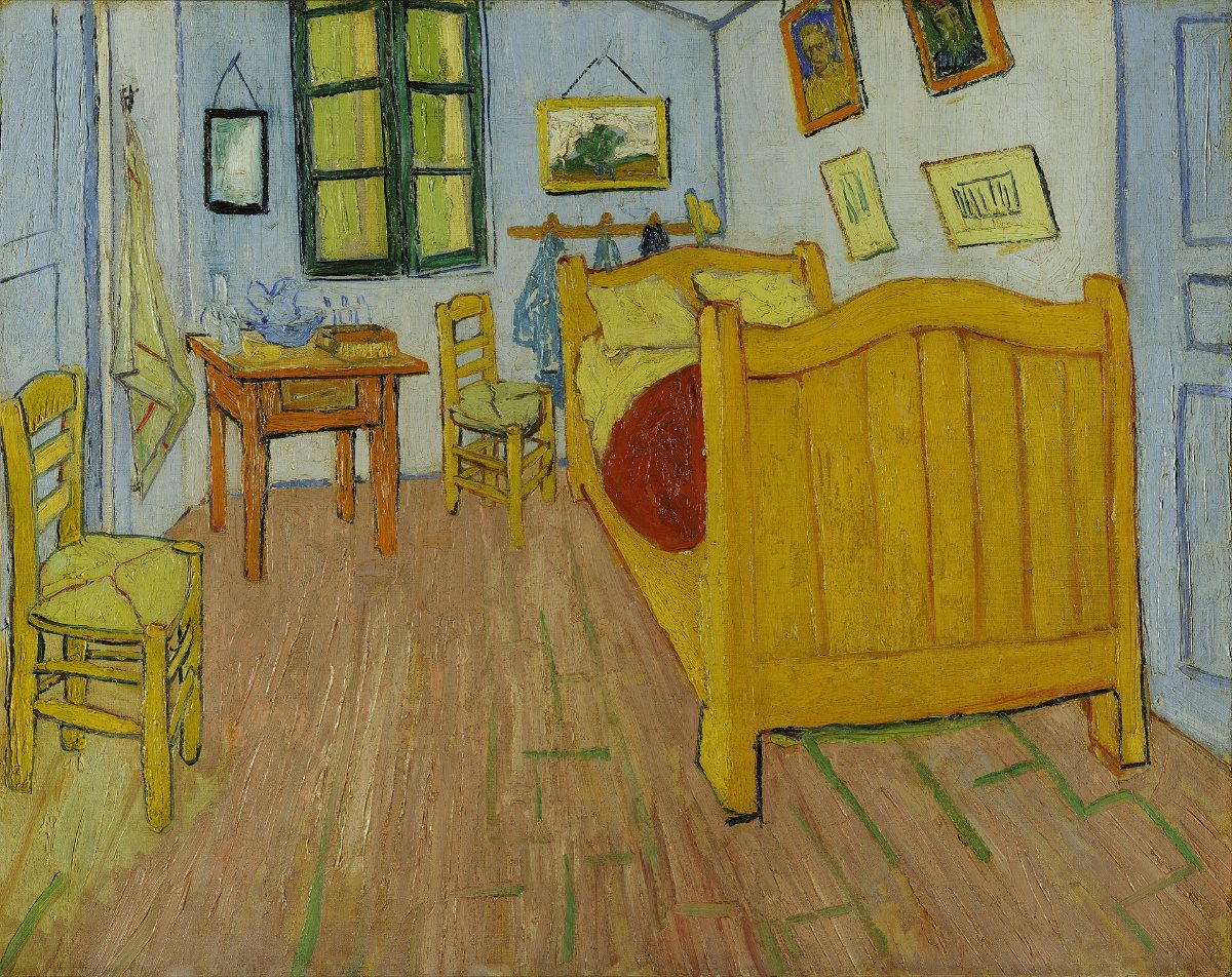
Vincent van Gogh's The Bedroom: a small room with a wooden bed, two chairs, a table, framed pictures on blue-toned walls, and a red-brown floor.
Vincent van Gogh, The Bedroom, 1889 · The painting as it appears today.
A conservator's gloved hand raises the carved edge of a historic frame by a few millimetres, revealing a narrow violet pigment line protected beside the more blue-looking exposed paint.
Concept visualization of a protected edge clue: less-exposed colour can survive beneath a frame lip. It illustrates the evidence principle, not a measured image of The Bedroom.

A reconstruction should show its uncertainty

Some conclusions are strongly supported by surviving pigment and chemical maps. Others depend on optical models, expert judgement, or incomplete clues. An honest digital reconstruction always says which parts were actually seen, which parts were figured out, and which parts are still a guess.
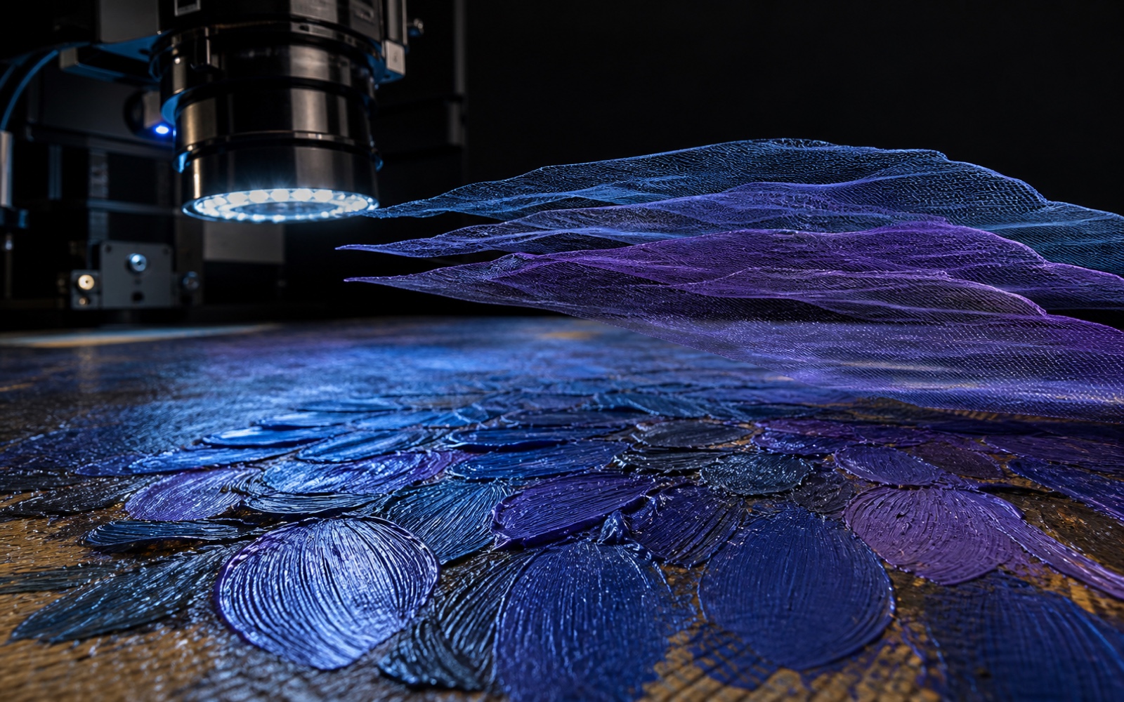
An intact impasto surface remains beneath a non-contact imaging lens while three closely spaced blue-to-violet digital meshes hover above the same registered brushstroke geometry.
Concept visualization: several closely related colour hypotheses remain registered to the same intact brushstrokes. Their separation represents model uncertainty; this is not a measured uncertainty map from Getty.

Look again

If the colours of a painting can change with time, what are we looking at today: only Van Gogh's work, or a work made jointly by Van Gogh and time?